

Autor :Saraguro RamĂrez, Byron L.1, Jaramillo, Byron1, Chico, Wendy1, MenĂ©ndez, Denisse2, Rueda, MarĂa JosĂ©3, LĂłpez, MarĂa Fernanda4
1 Hospital General Instituto Ecuatoriano de Seguridad Social IESS Babahoyo. Babahoyo-Ecuador 2 Hospital General Instituto Ecuatoriano de Seguridad Social IESS Quevedo. Quevedo-Ecuador 3 Facultad de EnfermerĂa Pontificia Universidad CatĂłlica del Ecuador. Quito-Ecuador 4Universidad Nacional de Chimborazo. Riobamba-Ecuador.
https://doi.org/10.56538/ramr.HCCY4307
Correspondencia : Byron Leonel Saraguro RaÂmĂrez - Ecuador. Province of Pichincha. CantĂłn RumiÂñahui. Parroquia San Pedro de Taboada. Gaspar Lema Street, 159 - E-mail: byronÂsaraguromd@gmail.com
Received: 11/13/2022
Accepted: 02/11/2023
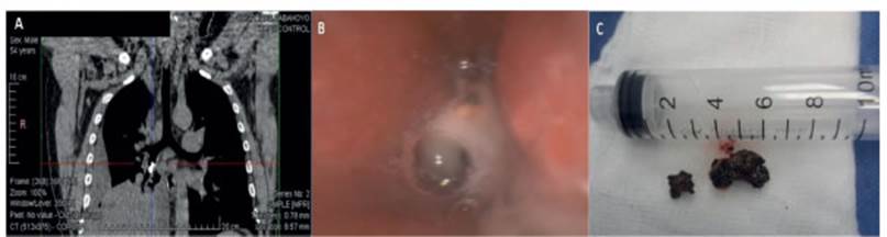
CASE 1
Male patient, 54
years old, with no personal medical record, presenting with a 15-day history of
cough accompanied by hemoptysis and dyspnea with a score of 2 according to the mMRC (Modified Medical Research Council) scale, with no
apparent cause. The patient was
referred to our Unit for suspected right pleural effusion. Chest tomography was
requested, revealing a hyperdense image at the level
of the intermediate bronchus and obstructive atelectasis of the middle lobe and
the right lower lobe. Foreign body extraction (stone) was performed using rigid
bronchoscopy (Figure 1).
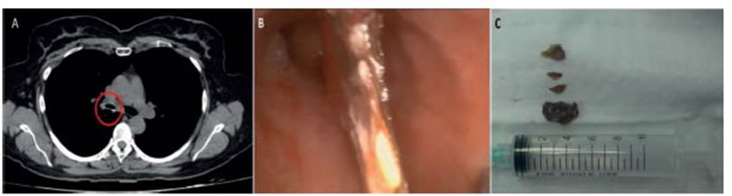
CASE 2
Female patient, 52 years old,
with a history of depressive syndrome, presenting with a one and a half-year
history of cough without expectoration. OccasionÂally she presented isolated
episodes of hemoptysis. She received treatment with short-acting
beta-adrenergic agonists (SABAs) suspected to have asthma, without relief of
the cough. Due to clinical disagreement, chest tomography was requested,
revealing a hyperdense image in the right main
bronchus. Foreign body extraction (fish bone) was performed using rigid
bronchoscopy.
Aspiration of foreign bodies is a
common accident in pediatrics. 75 % of the cases occur between the ages of two
and three, and 15 % in children over 6 years old.
It is an uncommon clinical entity
in adults and requires a high index of suspicion. It usually presents as acute
respiratory failure with nonspecific symptoms, such as chronic pneumonia,
atelectasis, chronic cough, or bronÂchospasm crises, without a history of bronchoaspiration.
In adults, it occurs after the
sixth decade of life, with risk factors such as neurodegenerative or
neuromuscular diseases that trigger abnormal airway protection mechanisms,
altered cough reflex, and dysphagia.
The foreign body appears in the
trachea in 4 % to 13 % of the cases, while bronchial localization ranges from
67 % to 80 %, with the right bronchus being more common, accounting for 52 % to
56 % of the cases.
Physical examination was normal
in 8 % to 10.4 % of the cases. Signs and symptoms depend on the nature, size,
location, and duration of the foreign body’s presence in the bronchial tree.
Radiological imaging is usually
normal in 9 % to 34 % of the cases. CT scans can help demonstrate the foreign
body, which may not always be visible on chest X-rays.
Rigid bronchoscopy is the
preferred therapeutic method for extracting a foreign body; however, it does
not constitute the gold standard. It allows for adequate airway protection and
ventilation, as well as better visualization of the object. It has a wider
working channel, allowing the passage of various types of forceps and grasping
instruments. It is performed under general anesthesia and can be used as a
backup method if flexible bronchoscopy fails. It is quicker and safer in
patients with respiÂratory failure, during episodes of asphyxia, in cases where
the foreign body is radiopaque, in unilateral hypoventilation, or obstructive
emphysema.1
Flexible bronchoscopy has proven
to be the best diagnostic method as it is easier, less expensive, and does not
require general anesthesia. The extraction of foreign bodies using a fiberoptic bronchoscopy is especially useful for those
lodged in smaller bronchi. Although it is not the ideal technique, it can
sometimes be used prior to rigid bronchoscopy to locate unclear foreign bodies.
It can also be introduced through the rigid bronÂchoscope as a combined
technique. Case series showing high efficacy of flexible bronchoscopy in
removing foreign bodies have been reported. The largest was the one reported by
Singh et al in a systematic review over a 35-year period with a sucÂcess rate
of 89.6 %, and by Ma et al, with a success rate of 73.7 % in 43 patients over a
thirteen-year period.2
Conflict of interest
Authors have no conflicts of
interest to declare.
REFERENCES
1.
Revuelta F, GarcĂa R, Pina I. Cuerpo extraño en vĂa aĂ©rea. Caso clĂnico y
revisiĂłn de literatura. Arch BronÂconeumol. 2020;56:395-408. https://doi.org/10.1016/j.arbres.2019.12.019
2. Ma W, Hu J, Yang M.
Application of flexible fiberoptic bronchoscopy in
the removal of adult airway foreing bodies. BMC
Surgery 2020;20:165. https://doi.org/10.1186/s12893-020-00825-5

| GalerĂa de imágenes | ||
| Mujer joven con afectaciĂłn pulmonar bilateral y alteraciĂłn de la conciencia | ||
Autores: Churin Lisandro |
 |
|